All published articles of this journal are available on ScienceDirect.

Isolation, Selection, and Identification of Phytophthora spp. Causing Pod Rot of Cocoa

Authors Info & Affiliations
Abstract
Introduction
Cocoa pod rot caused by Phytophthora spp. is a major constraint to cocoa production. This study aimed to isolate, characterize, and identify Phytophthora isolates associated with cocoa pod rot in Vinh Long province, Vietnam, to support disease-management strategies.
Methods
Twenty diseased cocoa pod samples were collected from 20 orchards in Phu Tuc and Tan Phu communes. Seven Phytophthora isolates were recovered and evaluated for colony growth, morphology, and pathogenicity. Four representative isolates were identified using ITS-rDNA sequencing and phylogenetic analysis.
Results
The isolates grew fastest on PCA medium, followed by V8 and PDA, with colony radii of 3.51–4.43, 2.93–3.99, and 2.03–3.42 cm, respectively, at 96 h after inoculation. All isolates produced papillate sporangia and terminal or intercalary chlamydospores and caused dark brown to black lesions on cocoa pods. Re-isolation fulfilled Koch’s postulates. PT10T was the most aggressive isolate, producing an 8.30-cm lesion diameter at 5 days after inoculation. The four sequenced isolates showed 100% ITS-rDNA similarity to P. palmivora and clustered with reference sequences of this species.
Discussion
Morphological, pathogenicity, and ITS-rDNA evidence supported identification of the sequenced isolates as P. palmivora. However, single-locus identification may not fully resolve closely related Phytophthora species.
Conclusion
P. palmivora was associated with cocoa pod rot in the surveyed orchards, with PT10T showing the greatest aggressiveness. These findings provide isolates for further studies on biological control and integrated disease management.
1. INTRODUCTION
Globally, the cultivated area of cocoa reaches approximately 11 million hectares, with an average yield of 480.5 kg ha-1. Cocoa production is an important source of income for millions of farmers in tropical regions, mainly in West Africa, Latin America, and Southeast Asia. In Vietnam, the cocoa production area is about 2,530 ha with a total production of approximately 1,260.84 tons and an average yield of 476.9 kg ha-1 [1, 2]. Cocoa is widely used for the production of chocolate, cocoa butter, and many other economically valuable products [3, 4]. In addition, cocoa beans provide numerous health benefits, including cardiovascular protection, neuroprotection, anti-inflammatory, and anticancer effects, mainly due to phenolic and methylxanthine compounds [5, 6]. Cocoa has also been shown to influence the composition of gut microbiota [7].
Black pod rot is the most serious disease affecting cocoa, causing significant economic losses with an estimated reduction of more than 30% of global cocoa production. In cocoa plantations, yield losses may reach up to 90% [8]. The disease usually begins as a small lesion on the pod surface, which rapidly expands to cover the entire pod, resulting in dark brown discoloration and gradual tissue necrosis [9, 10]. Phytophthora spp. primarily spread through water, where zoospores swim toward plant roots and tissues to infect the host, absorb nutrients, and secrete proteins that weaken host plants. At the same time, the pathogen forms spores and survival structures such as chlamydospores and oospores, enabling long-term survival and continuation of the infection cycle [11, 12].
The genus Phytophthora comprises many species infecting cocoa, the most common being P. palmivora, P. megakarya, P. citrophthora, and P. capsici, with different distributions depending on geographic regions and climatic conditions [13]. P. megakarya is the most destructive pathogen in Central and West Africa due to its high aggressiveness [14-16]. Under favorable natural conditions in Central and South America, P. capsici causes severe damage to cocoa plantations. Besides P. capsici and P. palmivora, two other species, P. citrophthora and P. heveae, have also been reported to cause serious damage in Brazil and Mexico [17-19]. In addition to pod rot, some species can infect stems, branches, and leaves, causing stem rot, branch dieback, and leaf blight [20-22].
Morphological characteristics are an important basis for species identification and classification. In the genus Phytophthora, morphological traits and growth characteristics on culture media are commonly used for species identification [23]. However, PCR techniques offer significant advantages over traditional methods due to their high sensitivity and flexibility [24]. The ITS (Internal Transcribed Spacer) region of rDNA has been widely used for identification and phylogenetic analysis of Phytophthora species [25-28].
Isolation and accurate identification of Phytophthora species are essential for understanding the biological characteristics of the pathogen. Morphology-based identification often faces limitations because species within this genus share very similar features. In addition, the evolution of a single gene does not necessarily represent the evolution of the entire genome [29]. Therefore, molecular analyses using gene regions such as ITS, COI, or β-tubulin are commonly employed for precise identification [30, 31]. Hence, this study was conducted to detect and identify Phytophthora species causing cocoa pod rot in Vinh Long province in order to develop effective disease management strategies.
2. MATERIALS AND METHODS
2.1. Materials
Phytophthora spp. isolates were obtained from 20 samples collected from cocoa orchards in Phu Tuc and Tan Phu communes, Vinh Long province, Vietnam.
Study period: from August 2025 to February 2026.
Water agar medium (WA): 15 g agar powder (produced by Hai Long Company, Vietnam) in 1 L distilled water.
Potato Dextrose Agar (PDA) medium: 39 g Potato Dextrose Agar (Merck, Germany) dissolved in 1 L distilled water.
V8 medium: 100 mL V8 Original vegetable juice (Campbell Soup Company, USA), 3 g CaCO3 (Xilong, Xinjiang, China), and 15 g agar powder dissolved in 1 L distilled water.
Potato Carrot Agar (PCA) medium: 20 g carrot, 20 g potato, and 15 g agar powder dissolved in 1 L distilled water.
2.2. Methods
2.2.1. Disease Sampling Method
In each orchard, diseased cocoa pods were randomly collected from five different positions and combined into one composite sample representing that orchard. The pods were cut using a knife sterilized with 70% ethanol and placed into separate plastic bags corresponding to each orchard. Before sampling, disease symptoms were recorded and photographed [32].
Location of the experiment: Microbiology Laboratory, Plant Morphology and Nutrition Laboratory, Faculty of Crop Science, College of Agriculture, Can Tho University.
2.2.2. Pathogen Isolation Method
After being brought to the laboratory, the cocoa pods were cleaned with 70% ethanol. The diseased tissue was then cut into small pieces with a diameter of 5 mm at the lesion margin between diseased and healthy tissues. The tissue pieces were immersed in 70% ethanol for 30 seconds and then dried with sterile blotting paper. The tissue pieces were subsequently placed onto WA medium. The isolation plates were incubated at 25 ± 2°C. Oomycete colonies were observed after 2 days. If colonies showed different morphological characteristics, the hyphal tips were transferred to plates containing 10 mL PDA medium to obtain pure cultures [32, 33]. The pure cultures were preserved in test tubes containing 20 mL sterile distilled water.
2.2.3. Characteristics of Pathogen
2.2.3.1. Colony Growth Rate
The experiment was arranged in a Completely Randomized Design (CRD). Each Phytophthora isolate represented one treatment with five replicates corresponding to five Petri dishes. Each oomycete isolate was cultured on Petri dishes containing V8 medium. A mycelial plug with a diameter of 5 mm was cut and placed in the center of the Petri dish. The plates were then incubated under alternating light and dark conditions (12 h light/12 h dark) at 25 ± 2°C.
The same procedure was conducted on PDA and PCA media. Colony color, colony morphology, and growth pattern on the media were observed after 5 days, and colony radius (cm) was measured at 48, 72, 96, 120, and 144 hours after inoculation (HAI). Measurements were discontinued when a colony reached the edge of the Petri dish.
2.2.3.2. Morphological Characteristics of Sporangia and Chlamydospores
Preparation of soil solution: One hundred grams of cocoa rhizosphere soil (sieved to 2 mm) was weighed, and distilled water was added to reach a total volume of 1 L. The mixture was stirred thoroughly, filtered, and stored in a glass bottle under cool conditions [33].
Preparation of 10% diluted soil solution: The soil solution was diluted at a ratio of 100 mL soil solution to 1 L distilled water and sterilized at 121°C for 20 minutes.
Preparation of oomycete samples: Phytophthora isolates were cultured on V8 medium in Petri dishes for 5–7 days under room conditions. Mycelial plugs with a diameter of 5 mm were then cut and transferred to new Petri dishes. Five milliliters of the diluted soil solution was added. The cultures were incubated under alternating light and dark conditions (12 h light/12 h dark) at 25 ± 2°C for 5 days. Different types of spores were observed at the margins of the mycelial plugs [33]. The shape and size of chlamydospores and sporangia, as well as the papillation of the sporangia, were observed and measured under a 40X objective lens [34].
2.2.3.3. Pathogenicity
The experiment was arranged in a CRD. Each Phytophthora spp. isolate represented one treatment with three replicates corresponding to three cocoa pods, a oomycete slice per one cocoa pod. In the control treatment, a slice of PDA medium was used.
Artificial inoculation following Koch’s postulates: Healthy cocoa pods were surface-sterilized with 70% ethanol. One pod was placed in a box and maintained under moist conditions using sterile blotting paper supplemented with 5 mL sterile distilled water. A wound was made on the pod surface, and a 5 mm mycelial plug was placed onto the wound. The same procedure was performed for the control treatment, but only a PDA plug was applied [32]. After disease symptoms appeared, the pathogen was re-isolated from the diseased tissues and compared with the original isolate.
Twenty μL of sterile distilled water was added to the mycelial plug daily. Lesion diameter (cm) was determined at 2, 3, 4, and 5 days after inoculation (DAI).
2.2.4. Identification of Phytophthora spp.
Phytophthora spp. isolates were cultured in 15 mL of V8 medium in the dark at 25 ± 2°C for 5 days for DNA extraction. PCR amplification of the ITS-rDNA region was performed using the primers ITS1 (5′-TCCGTAGGTAACCTGCGG-3′) and ITS4 (5′-TCCTCCGCTTATTGATATGC-3′) according to White et al. [35].
The PCR cycling conditions for the ITS rDNA region were as follows: for the ITS rDNA region, the PCR reaction was initiated with an initial denaturation at 95°C for 5 minutes, followed by 35 cycles consisting of denaturation at 95°C for 1 minute, annealing at 54°C for 30 seconds, and extension at 72°C for 1 minute. The reaction was completed with a final extension at 72°C for 5 minutes and then held at 4°C.
The amplicons were purified using the TIANquick Midi purification kit. The purified products were verified by 1.0% (w/v) agarose gel electrophoresis under UV light. The PCR products were subsequently sequenced using the Macrogen DNA sequencing service. The obtained sequences were analyzed using BioEdit version 7.0.5.3 and ChromasPro version 1.7, and then compared with sequences available in the GenBank database using BLAST on NCBI. The phylogenetic tree was constructed using the Neighbor-Joining method in MEGA version 11. The reliability of the inferred phylogenetic relationships was evaluated using 1,000 bootstrap replicates, and the bootstrap support values are shown next to the branches.
2.2.5. Phylogenetic Analysis
Phylogenetic tree construction: The Clustal-W program was used to align nucleotide sequences and identify differences among gene regions of the genera and species. The phylogenetic tree illustrating genetic relationships among species was constructed using MEGA version 11.
3. RESULTS
3.1. Collection and Isolation of Phytophthora spp. Causing Cocoa Pod Rot
Cocoa pods showing pod rot symptoms were collected. The symptoms appeared as dark brown, water-soaked lesions occurring at the pod peduncle (Fig. 1a), the middle part of the pod, or the pod tip (Fig. 1b). The lesions gradually turned black and covered the entire surface of the pod husk. In addition, the pathogen was also observed to infect other vegetative parts of the plant, showing symptoms such as leaf blight (Fig. 1c), leaf yellowing (Fig. 1d), and branch dieback on mature trees (Fig. 1e).
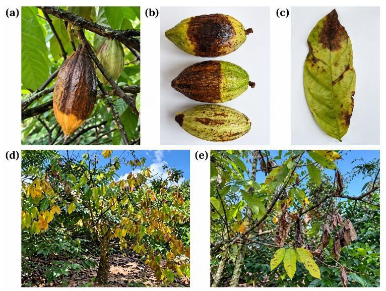
Disease symptoms caused by Phytophthora spp. on cocoa plants. (a) Cocoa pod rot on the tree; (b) Cocoa pod showing black rot with expanding lesions; (c) Leaf blight symptoms with brown necrotic lesions; (d) Cocoa tree showing severe leaf yellowing; (e) Branch dieback and canopy wilting symptoms.
A total of seven Phytophthora spp. isolates were obtained from 20 diseased cocoa pod samples collected in Vinh Long province (Fig. S1). All seven isolates were used to observe morphological characteristics, colony morphology, growth rate, and pathogenicity on cocoa pods.
3.2. Morphological Characteristics and Colony Morphology of Phytophthora spp. Causing Cocoa Pod Rot
3.2.1. Colony Morphology
On V8 medium, all isolates produced white, velvety, fluffy, or stellate mycelia with a cottony texture, forming concentric or irregular patterns and spreading radially from the center (Fig. 2); (Fig. S2).
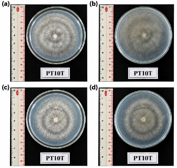
Colony morphology of Phytophthora spp. isolate PT10T cultured at 25 ± 2°C. (a, b) Upper and reverse sides of colonies on V8 medium at 120 HAI; (c, d) Upper and reverse sides of colonies on PDA medium at 144 HAI.
3.2.2. Spore Characteristics
Based on morphology, the oomycete isolates obtained from cocoa pods exhibited typical characteristics of Phytophthora spp. The chlamydospores ranged 25–53 µm in size and were formed terminally or intercalarily on the hyphae (Table 1). The sporangia were papillate, with shapes ranging from pyriform (pear-shaped) and ovoid to irregular, with lengths of 20–63 µm and widths of 10–58 µm (Table 1, Fig. 3). The sporangial structures were polymorphic in shape (pyriform, ovoid, or irregular) (Table 1). The morphological similarity among the isolates suggests that the isolates belong to the same species commonly causing disease on cocoa.
| Isolate | Chlamydospore | Sporangium | ||||
|---|---|---|---|---|---|---|
| Size (µm) | Formation Position | Shape | Length (µm) | Width (µm) | Papilla | |
| PT1T | 25–50 | Terminal, intercalary | Pyriform, irregular, ovoid | 25–55 | 15–38 | Present |
| PD6T | 29–48 | Terminal, intercalary | Pyriform, irregular, ovoid | 20–43 | 10–30 | Present |
| PT7T | 30–50 | Terminal, intercalary | Pyriform, irregular, ovoid, spherical | 30–63 | 20–40 | Present |
| PT9T | 26–45 | Terminal, intercalary | Pyriform, irregular, ovoid | 30–65 | 23–40 | Present |
| PT10T | 33–46 | Terminal, intercalary | Pyriform, irregular, ovoid, spherical | 38–63 | 20–40 | Present |
| PT11T | 33–50 | Terminal, intercalary | Pyriform, irregular, ovoid | 28–63 | 15–58 | Present |
| PT12T | 36–53 | Terminal, intercalary | Pyriform, irregular, ovoid | 28–50 | 18–33 | Present |
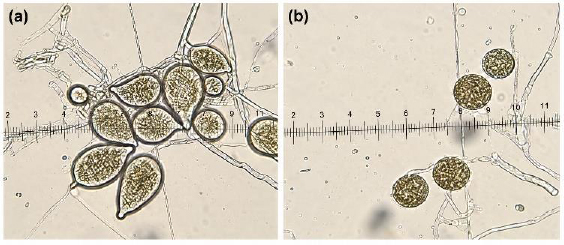
Spore characteristics of Phytophthora sp. isolate PT10T observed under a light microscope (40X objective), including: (a) Sporangium; (b) Chlamydospore.
3.3. Colony Growth Rate
The Phytophthora spp. isolates showed good growth on all three agar media, including PCA, PDA, and V8. On PCA medium, Phytophthora spp. exhibited the fastest growth, followed by V8 medium. Under cultivation on PDA medium, colonies of Phytophthora spp. reached their maximum size at 120 HAI (Table 2).
| Medium | Isolate | 48 | 72 | 96 | 120 | 144 |
| Hours | ||||||
| PCA | PT1T | 1.32c | 2.33e | 3.51d | - | - |
| PT7T | 1.63b | 2.92b | 4.33a | - | - | |
| PT9T | 1.52b | 2.82bc | 4.13b | - | - | |
| PT10T | 1.52b | 2.77cd | 4.37a | - | - | |
| PT11T | 1.50b | 2.68d | 3.72c | - | - | |
| PT12T | 1.67a | 3.08a | 4.43a | - | - | |
| PD6T | 1.52b | 2.72cd | 4.37a | - | - | |
| CV (%) | 4.64 | 2.56 | 2.54 | - | - | |
| Significance | * | * | * | |||
| PDA | PT1T | 0.89c | 1.67e | 2.13d | 3.00e | 4.29b |
| PT7T | 1.09b | 1.84d | 2.47c | 3.42d | 4.43ab | |
| PT9T | 1.05b | 1.86d | 2.88b | 3.90c | 4.50a | |
| PT10T | 1.22a | 2.47a | 3.42a | 4.45a | 4.38ab | |
| PT11T | 1.03b | 1.42f | 2.03d | 2.75f | 3.95c | |
| PT12T | 1.20a | 2.29b | 2.43c | 4.05b | 4.50a | |
| PD6T | 1.00b | 2.05c | 2.90b | 3.42d | 4.37ab | |
| CV (%) | 6.62 | 4.31 | 2.97 | 1.77 | 2.72 | |
| Significance | * | * | * | * | * | |
| V8 | PT1T | 1.15c | 2.13c | 2.93e | 4.00b | - |
| PT7T | 1.23bc | 2.38b | 3.54c | 4.50a | - | |
| PT9T | 1.32b | 2.40b | 3.45cd | 4.50a | - | |
| PT10T | 1.51a | 2.73a | 3.99a | 4.50a | - | |
| PT11T | 1.20c | 2.12c | 2.98e | 3.94b | - | |
| PT12T | 1.47a | 2.75a | 3.84b | 4.50a | - | |
| PD6T | 1.53a | 2.43b | 3.34d | 4.50a | - | |
| CV (%) | 4.70 | 2.92 | 2.76 | 0.99 | - | |
| Significance | * | * | * | * | - | |
The structures of the different spore types observed under a light microscope (40X objective) are clearly illustrated in Fig. (3).
On PCA medium, at 96 HAI, the colony radii of the isolates ranged from 3.51 cm to 4.43 cm. Among them, isolates PT12T, PT10T, PD6T, and PT7T showed rapid growth (4.33–4.43 cm). In contrast, isolate PT1T exhibited the slowest growth, reaching 3.51 cm (Table 2).
On PDA medium, at 144 HAI, the colony radius of the isolates ranged from 3.95 to 4.50 cm. Isolates PT9T and PT12T showed good adaptation and growth, reaching the maximum size of 4.50 cm. Isolate PT11T showed the slowest growth on PDA medium, with a size of 3.95 cm (Table 2).
On V8 medium, the isolates grew rapidly and uniformly. At 120 HAI, isolates PT7T, PT9T, PT10T, PT12T, and PD6T reached the maximum colony size of 4.50 cm. In contrast, isolates PT1T and PT11T exhibited slower growth, ranging from 3.94 to 4.00 cm (Table 2).
3.4. Pathogenicity of Phytophthora spp.
The lesion diameter increased progressively over time in all treatments. At 2 DAI, isolates PT12T and PT10T showed high pathogenicity, with lesion diameters of 1.95 cm and 1.90 cm, respectively, whereas isolate PT1T produced the smallest lesion diameter (0.627 cm). At 3 and 4 DAI, isolate PT12T exhibited the fastest lesion expansion, with lesion diameters of 4.03 cm and 6.03 cm, respectively. At 5 DAI, isolate PT10T showed the highest pathogenicity, reaching a lesion diameter of 8.3 cm. In contrast, isolates PT1T and PT11T produced the smallest lesions, measuring 6.53 cm and 6.66 cm, respectively (Fig. 4).
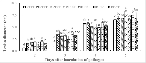
Lesion development caused by seven Phytophthora isolates on detached cocoa pods at 2, 3, 4, and 5 days after inoculation. Note: Mean values within the same bars followed by the same lowercase letter are not significantly different according to Duncan’s multiple range test.
The typical symptoms observed were dark brown to black necrotic lesions, accompanied by the abundant development of fine white mycelia on the pod surface (Fig. 5).
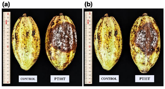
Pathogenicity of Phytophthora spp. PT10T (high pathogenicity) and PT11T (low pathogenicity) on cocoa pods at 5 DAI. Based on the growth and pathogenicity of Phytophthora spp. isolates under different media, 4 isolates (PD6T, PT7T, PT10T, and PT12T) were selected for identification.
The Phytophthora spp. re-isolated from artificially induced lesions on cocoa pods exhibited morphological characteristics consistent with the original isolates, thereby fulfilling Koch’s postulates.
All seven isolates were included in the morphological, cultural, and pathogenicity assessments. Based on their combined colony-growth and pathogenicity characteristics, four isolates, PD6T, PT7T, PT10T, and PT12T, were selected as representative isolates for ITS-rDNA sequencing. These isolates represented the observed variation in growth and lesion development among the recovered isolates. The remaining isolates were retained in the phenotypic analyses but were not included in the molecular analysis.
3.5. Identification of Phytophthora spp. Isolates Causing Cocoa Pod Rot
All selected oomycete isolates showed 100% sequence similarity with P. palmivora. In the phylogenetic tree, four isolates (PD6T, PT7T, PT10T, and PT12T) exhibited different levels of virulence, and all clustered within the same clade as P. palmivora strains available in GenBank (Fig. 6). Among them, isolate PT10T, which showed the highest virulence and fastest growth rate, was selected for subsequent experiments (Table 2, Fig. 5).
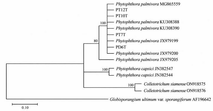
Phylogenetic tree based on ITS gene sequences of Phytophthora spp. isolates obtained from cocoa. The tree was reconstructed using the Neighbor-Joining method in MEGA version 11. Branch support was evaluated using 1,000 bootstrap replicates, and bootstrap values ≥50% are shown at the nodes. Globisporangium ultimum var. sporangiiferum AF196642 was used as the outgroup. The scale bar represents 0.10 substitutions per nucleotide position.
4. DISCUSSION
Isolation results from diseased cocoa pod samples collected in Vinh Long province yielded seven oomycete isolates exhibiting the typical characteristics of P. palmivora (Table 1, Fig. S1). The colony characteristics and morphological features were consistent with the standard description of P. palmivora reported by Saul Maora et al. [36]. All isolated Phytophthora strains were able to produce chlamydospores, with sizes ranging from 25–53 µm (Table 1). The sporangia were papillate, with shapes varying from pyriform and ovoid to irregular, with lengths ranging from 20–63 µm and widths from 10–58 µm (Fig. 3).
The Phytophthora isolates were able to grow on various culture media, including V8, PDA, and PCA (Table 2). Koch’s postulates confirmed that all seven isolates caused typical cocoa pod rot symptoms, characterized by expanding dark brown lesions (Fig. 5). This is consistent with Debnath et al. [37]. At 7 DAI, the lesions became darker and larger, accompanied by the appearance of abundant mycelia and spores. P. palmivora strains showed a growth rate of 3.8 cm within 2 days on V8 medium. These results indicate that P. palmivora isolated from cocoa pods collected in Vinh Long exhibits high virulence, particularly isolate PT10T, which produced lesions of 8.3 cm after 5 days of artificial inoculation (Table 2). The identification results and pathogenicity evaluation are consistent with the current disease situation in Vietnam. According to Hoa et al. [38], in the Southeast, Central Highlands, and Southwest regions of Vietnam, all cocoa-growing areas have been affected by black pod disease caused by P. palmivora. Puig [39] also reported P. palmivora causing cocoa pod rot in Puerto Rico. Similar findings were reported by Maizatul-Suriza et al. [40] worldwide.
The ITS-rDNA sequences of the four selected isolates showed 100% similarity to P. palmivora reference sequences and clustered with this species in the phylogenetic analysis. However, ITS is a single locus and may not adequately distinguish closely related Phytophthora species or reflect genome-wide evolutionary relationships [41]. Therefore, future studies should combine ITS with additional mitochondrial and nuclear loci, such as COI or COII and β-tubulin, and should include a wider range of reference sequences and isolates from different cocoa-growing regions [30].
Phytophthora palmivora is widely distributed in cocoa-producing regions and can infect pods, stems, leaves, and other plant tissues. It has been reported to cause 20–30% pod losses and cause the death of up to 10% of cocoa trees annually through stem cankers [42]. The pathogen has also been associated with diseases of economically important perennial crops, including durian, rubber, and oil palm [43-45]. The recovery of aggressive P. palmivora isolates from Vinh Long province emphasizes the need for continued pathogen monitoring and the development of locally appropriate disease-management strategies.
STUDY LIMITATIONS
This study had several limitations. First, the samples were collected from only two communes in Vinh Long province, and the seven recovered isolates may not represent the full phenotypic and genetic diversity of Phytophthora populations in other cocoa-growing regions of Vietnam. Second, pathogenicity was evaluated using detached cocoa pods under controlled laboratory conditions. This assay may not fully represent disease development under field conditions, where temperature, humidity, host developmental stage, inoculum pressure, and interactions with other microorganisms may influence infection. Third, only four representative isolates were subjected to molecular identification, while the remaining three isolates were characterized based on morphology, growth, and pathogenicity. Finally, molecular identification and phylogenetic analysis were based only on the ITS-rDNA region. Because a single locus may not adequately resolve closely related Phytophthora taxa or represent genome-wide relationships, future studies should use broader geographical sampling, field-based pathogenicity assessments, and multilocus analyses incorporating markers such as COI, COII, and β-tubulin.
CONCLUSION
One isolate was selected from seven Phytophthora spp. isolates causing cocoa pod rot. Based on colony morphology, spore size and morphology (including chlamydospores and sporangia), combined with molecular identification, the pathogen was identified as Phytophthora palmivora isolate PT10T, showing 100% sequence similarity. Isolate PT10T exhibited a growth rate of 3.99 cm at 96 HAI on V8 medium and produced a lesion diameter of 8.3 cm at 5 DAI. However, this study was limited by the small number of Phytophthora isolates obtained from cocoa orchards in only two communes of Vinh Long province; therefore, the results may not fully represent the diversity of Phytophthora populations in other cocoa-growing regions. However, the study was limited to a small sampling area, detached-pod pathogenicity assays, and identification using a single molecular locus. Future studies should include broader geographical sampling, field-based pathogenicity evaluations, and multilocus analyses using markers such as COI, COII, and β-tubulin to improve taxonomic resolution and characterize the genetic diversity of P. palmivora populations in Vietnam.
AUTHORS’ CONTRIBUTIONS
The authors confirm their contribution to the paper as follows: H.T.Q., L.T.Q., and N.Q.K.: Data collection; H.T.Q., N.D.T., L.T.M.T., N.T.T., and L.T.T.: Analysis and interpretation of results; H.T.Q., L.T.Q., L.T.T., and N.Q.K.: Draft manuscript preparation; H.T.Q., L.T.Q., and N.Q.K. All authors reviewed the results and approved the final version of the manuscript.
LIST OF ABBREVIATIONS
| ANOVA | = Analysis of variance |
| BLAST | = Basic Local Alignment Search Tool |
| COI | = Cytochrome c oxidase subunit I |
| COII | = Cytochrome c oxidase subunit II |
| CRD | = Completely randomized design |
| CV | = Coefficient of variation |
| DAI | = Days after inoculation |
| DNA | = Deoxyribonucleic acid |
| HAI | = Hours after inoculation |
| ITS | = Internal transcribed spacer |
| ITS-rDNA | = Internal transcribed spacer region of ribosomal DNA |
| MEGA | = Molecular Evolutionary Genetics Analysis |
| NCBI | = National Center for Biotechnology Information |
| PCA | = Potato carrot agar |
| PCR | = Polymerase chain reaction |
| PDA | = Potato dextrose agar |
| SD | = Standard deviation |
| UV | = Ultraviolet |
| V8 | = V8 juice agar medium |
| WA | = Water agar |
AVAILABILITY OF DATA AND MATERIAL
All data generated or analyzed during this study are included in this published article.
ACKNOWLEDGMENTS
Declared none.